How Wiggly Proteins Guard the Genome
Dynamic network in the pores of the nuclear envelope blocks dangerous invaders
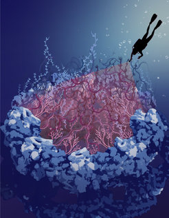
Tiny pores in the cell nucleus play an essential role for healthy aging by protecting and preserving the genetic material. A team in Germany from the Department of Theoretical Biophysics at the Max Planck Institute of Biophysics in Frankfurt am Main and the Synthetic Biophysics of Protein Disorder Group at Johannes Gutenberg University Mainz has literally filled a hole in the understanding of the structure and function of these nuclear pores. The scientists found out how intrinsically disordered proteins in the center of the pore can form a spaghetti-like mobile barrier that is permeable for important cellular factors but blocks viruses or other pathogens.
Text: Katharina Kaefer
Human cells shield their genetic material inside the cell nucleus, protected by the nuclear membrane. As the control center of the cell, the nucleus must be able to exchange important messenger molecules, metabolites or proteins with the rest of the cell. About 2000 pores are therefore built into the nuclear membrane, each consisting of about 1000 proteins.
For decades, researchers have been fascinated by the three-dimensional structure and function of these nuclear pores, which act as guardians of the genome: substances that are required for controlling the cell are allowed to pass, while pathogens or other DNA-damaging substances are blocked from entry. The nuclear pores can therefore be thought of as molecular bouncers, each checking many thousands of visitors per second. Only those who have an entrance ticket are allowed to pass.
How do the nuclear pores manage this enormous task? About 300 proteins attached to the pore scaffold protrude deep into the central opening like tentacles. Until now, researchers did not know how these tentacles are arranged and how they repel intruders. This is because these proteins are intrinsically disordered and lack a defined three-dimensional structure. They are flexible and continuously moving – like spaghetti in boiling water.

Combination of Microscopy and Computer Simulations
As these intrinsically disordered proteins (IDPs) are constantly changing their structure, it is difficult for scientists to decipher their three-dimensional architecture and their function. Most experimental techniques that researchers use to image proteins only work with a defined 3D structure. So far, the central region of the nuclear pore has been represented as a hole because it was not possible to determine the organization of the IDPs in the opening.
The team led by Gerhard Hummer, Director at the Max Planck Institute of Biophysics, and Edward Lemke, Professor for Synthetic Biophysics at Johannes Gutenberg University Mainz, and Adjunct Director at the Institute of Molecular Biology Mainz has now used a novel combination of synthetic biology, multidimensional fluorescence microscopy and computer-based simulations to study nuclear pore IDPs in living cells.
“We used modern precision tools to mark several points of the spaghetti-like proteins with fluorescent dyes that we excite by light and visualize in the microscope,” Lemke explains. “Based on the glow patterns and duration, we were able to deduce how the proteins must be arranged.” Hummer adds, “We then used molecular dynamics simulations to calculate how the IDPs are spatially organized in the pore, how they interact with each other and how they move. For the first time, we could visualize the gate to the control center of human cells.”
Dynamic Protein Network as Transport Barrier
The tentacles in the transport pore take on a completely different behavior compared to what we knew before, because they interact with each other and with the cargo. They move permanently like the aforementioned spaghetti in boiling water. So, in the center of the pore there is no hole, but a shield of wiggly, spaghetti-like molecules. Viruses or bacteria are too big to get through this sieve. However, other large cellular molecules needed in the nucleus can pass as they carry very specific signals. Such molecules have an entry ticket, whereas pathogens usually do not. “By disentangling the pore filling, we enter a new phase in nuclear transport research,” adds Martin Beck, collaborator and colleague at the Max Planck Institute of Biophysics.
"Understanding how the pores transport or block cargo will help us identify errors. After all, some viruses manage to enter the cell nucleus despite the barrier," Hummer sums up. “With our combination of methods, we can now study IDPs in more detail to find why they are indispensable for certain cellular functions, despite being error-prone. In fact, IDPs are found in almost all species, although they carry the risk of forming aggregates during the aging process which can lead to neurodegenerative diseases such as Alzheimer's,” Lemke says. By learning how IDPs function, researchers aim to develop new drugs or vaccines that prevent viral infections and help healthy aging.


