Werner Kühlbrandt – Structural Biology
How can we make the most of cryo-electron microscopy for determining the structure and function of membrane protein complexes?
The main aim of the Department is to understand the structure and function of membrane proteins and large membrane protein complexes as determined by electron cryo-microscopy (cryoEM) and X-ray crystallography. CryoEM of macromolecular assemblies is undergoing a revolution in resolution (Kühlbrandt, Science 2014), largely due to the development of new direct electron detectors, to which the Department and the Max Planck Society have contributed.
Membrane proteins play crucial roles in virtually all biomedical processes. It is therefore essential to understand exactly how they work. This requires structures at atomic or near-atomic resolution. To obtain these structures is challenging for several reasons: Many membrane proteins are fragile and tend to denature upon isolation; complexes can rarely be overexpressed or reconstituted, because they consist of many different membrane protein components; membrane proteins and especially membrane protein complexes are difficult to crystallize.
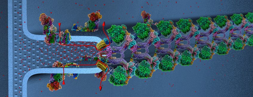
CryoEM is ideal for investigating membrane protein complexes, because this method does not depend on crystals. Samples are studied either in detergent (or amphipol) solution by single-particle cryoEM, or directly in the membrane by electron cryo-tomography (cryoET). Single-particle cryoEM has recently achieved resolutions that rival or even surpass X-ray crystallography.
"Resolution revolution". The years since 2014 have been a particularly stimulating and exciting time for cryoEM in general and for the Department in particular. We have considerably expanded the scope of membrane proteins and membrane systems we investigated during this period, not least in response to the numerous requests for promising collaborations from leading groups in membrane protein research well beyond Frankfurt and Germany. All in all, the past few years have been our most productive period to date. So far we have determined the structures of 18 different membrane protein assemblies by single-particle cryoEM at 2.7 to 9 Å, and of 15 by cryoET and subtomogram averaging at 18 to 30 Å resolution. The structures we determined have led to a plethora of fascinating and unexpected insights. In the next three to five years, we will take the membrane proteins and protein complexes we currently investigate to higher, hopefully near-atomic resolution, making use of the outstanding inhouse cryoEM equipment that Max Planck funding has enabled us to build up. This will provide us with a yet more detailed understanding of their molecular mechanisms.