Cellular Decoding Machines in Action
Researchers observe structural dynamics of ribosomes and their interactions with an anticancer drug while they translate the genetic code into proteins
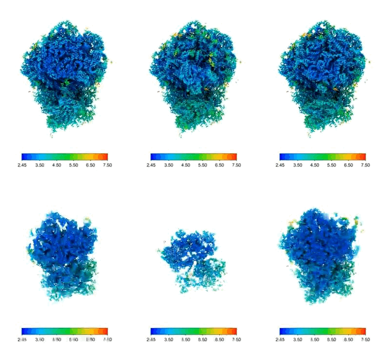
Scientists from the Department of Molecular Sociology at the Max Planck Institute of Biophysics have visualized the dynamic structural changes of ribosomes during protein production in human cells for the first time. Using high-resolution cellular tomography, they generated 3D images of ribosomes in action and thereby also deciphered the target site and mode of action of a leukemia drug that inhibits protein biosynthesis in cells.
Text: Katharina Kaefer
Every second, our cells produce proteins. Our body needs these proteins for all vital processes such as growth, metabolism or immune defense. Ribosomes are responsible for the assembly line production based on a strict blueprint. Like decoding machines, they translate a copy of the genetic code into a chain of amino acid building blocks at high speed that is later folded to the ready-to-use protein. During this process called translation, the ribosomes, which consist of RNA and proteins, undergo dynamic structural changes until they are finally recycled.

Interplay of Multiple Cellular Machines
The complete understanding of such biochemical processes including structure determination often contributes to a better understanding of physiological dysfunction or disease. In vitro, which means in the test tube, scientists have already studied the structure of ribosomes in the different translation stages. A team of researchers led by Martin Beck, Director at the Max Planck Institute of Biophysics, has now visualized the structural dynamics of ribosomes during translation in human cells at high resolution for the first time. They published their results in the journal Science.
"If we want to understand in detail how molecular machines, for example ribosomes, work and why their function is sometimes impaired, we should not just look at them in isolation," explains Martin Beck. "We need to study them in action in their natural environment and consider the interplay with other cell components." One can imagine it like with machines that we know from our everyday life. If the WiFi router does not work, it is not necessarily the device itself that is broken. Perhaps the power supply is disrupted, or a cable or a wire is damaged.
3D Images Created by Cellular Tomography


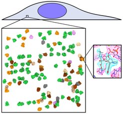
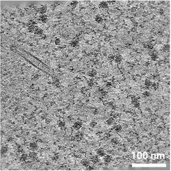
Beck's team generated 3D images of ribosomes at different stages in the cell's translational cycle using cellular tomography. "With state-of-the-art electron microscopes and sophisticated image processing, we can visualize ribosomes at near-atomic resolution inside human cells. To do so, we freeze whole cells and cut them into ultrathin slices", explains Huaipeng Xing, first author of the study. "An electron beam passes through the slices in the microscope. The image results from the interactions of the electrons with the different sample components. We take images from many different angles and process them to obtain 3D structures of the ribosomes in the cell."
Using high-resolution electron tomography, the researchers were not only able to image ribosomes in human cells, but also visualize how the leukemia drug homoharringtonine (HHT) binds to ribosomes. It consequently inhibits the translation - and thus the protein production - and makes ribosomes hibernate. "Imaging drug molecules at their site of action at high resolution could help to tailor them even more precisely to improve the effect and reduce side effects," concludes Beck.


