Study unravels details of autophagy machinery assembly
In vitro reconstitution of early events in autophagy
Researchers from the University of Vienna, the Max Planck Institute for Biology of Ageing in Cologne and the Max Planck Institute of Biophysics in Frankfurt am Main succeeded in the reconstitution of the autophagosome nucleation using recombinant components from yeast. In addition, a model of an Atg9 vesicle was constructed that contains all of the key components of lipids and proteins that are crucial for the early phase of autophagy.
Autophagy

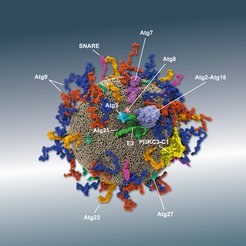
Model of an Atg9 vesicle from experimental data and bioinformatics predictions. The interaction of various Atg proteins and their partners enables Atg8 to be anchored in the membrane - an essential step for the development of the phagophore.
Even in the small units of life, the cells, savings are made: Autophagy is a natural regulatory mechanism of the cell, which ensures that unnecessary or defective components are dismantled recycled. In autophagy, cytoplasmic components (like mitochondria) are engulfed by a growing double membrane termed phagophore. Upon closure, this structure (now called autophagosome) isolates its cargo from the rest of the cell within a double-membraned vesicle. It fuses with a lysosome to start the process of waste management and disposal. The contents of the vesicle are then degraded and recycled.
Atg9 vesicles
It has long been known that vesicles with Atg9 protein (“Atg” autophagy-related) in their membrane play an essential role in autophagy, but the exact role is still unclear. The researchers show that Atg9 vesicles can serve as a platform for the formation of the phagophore. For the phagophore to grow, many Atg8 proteins are anchored in the growing membrane following a complicated sequence of events involving many proteins. In this work, a long sequence of events up to Atg8 anchoring was reconstituted in vitro with previously isolated proteins. This alloweds a precise view of the processes and shows new dependency relationships between the Atg proteins.
The growing phagophore double membrane needs a continuous supply of lipids. But where do they come from? One possibility is now shown: "Atg9 vesicles locate to the immediate vicinity of the endoplasmic reticulum (ER). In our model, the ER and Atg9 vesicles are virtually "short-circuited" by another protein, Atg2, so that lipids migrate from the ER to the growing vesicle. In this way the phagophore would be supplied with lipids from the ER", says Verena Baumann, one of the lead authors of the study in the laboratory of Sascha Martens from the Department of Biochemistry and Cell Biology at the Max Perutz Labs, University of Vienna.
Informative model observations
The reconstitution was also investigated by Sören von Bülow from the Max Planck Institute of Biophysics. In the Department of Theoretical Biophysics, led by Gerhard Hummer, a model of an Atg9 vesicle was built that contains all of the key components of lipids and proteins that are crucial for the early phase of autophagy. Von Bülow developed in-house analysis tools to show that the surface of the vesicle is tightly packed with proteins: "We postulate that the influx of lipids (from the ER) via Atg2 creates enough space on the membrane so that additional Atg8 can be anchored." Since defects in autophagy are associated with severe pathologies such as neurodegeneration, cancer, and infections, deciphering the mechanism is of the utmost importance for medicine.