Cryo-electron tomography

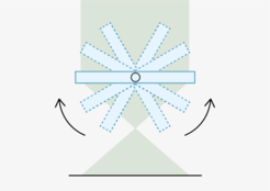
Cryo-electron tomography (CET) can provide 3D information of a single specimen, embedded in vitreous ice. This is achieved by tilting the sample in the microscope relative to the electron beam, and collecting sample images from many angles. The information of a tilt series is computationally combined to calculate a 3D volume of the sample, a so-called tomogram. Whereas for SPA, biological material is taken out of its native environment and studied in vitro, CET can provide structural information of the unaltered biological material in its biological context. Higher resolution of structures or proteins present in the tomogram can be achieved by sub-tomogram averaging, i.e. by combining the information from identical particles present in one or many tomograms. Automated data collection is performed on one of our 3 Titan Krios microscopes, equipped with energy filters K3, Falcon 3 or Falcon 4direct electron detectors.
One caveat of CET is that biological samples such as cells are typically too thick to be imaged in a TEM. It is therefore beneficial to combine the CET workflow with cryoFIB-SEM for lamella preparation (see below).