Cryo correlative light and electron microscopy

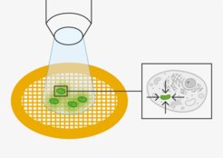
Fluorescence microscopy (FM) and electron microscopy (EM) are highly complementary techniques, and it can be beneficial to combine the information of both techniques from the same sample. This method is called correlative light- and electron microscopy (CLEM). Using fluorescence microscopy, regions of interest such as transfected cells, labelled proteins or rare events can easily be identified and mapped at relatively low magnification. The same regions of interest can then be targeted and examined at high resolution available with cryoEM. To perform cryo-fluorescence microscopy, a special cryostage is needed. We utilise the Leica EM cryoCLEM microscope, which combines widefield imaging with the SP8 confocal system. In the cryoCLEM workflow, cryo-fluoresence microscopy is typically followed by cryo-tomography with or without the FIB-milling of lamella.