Advanced Microscopy Tools
1. Superresolution Microscopy
2. Single Particle Stoichiometry Analysis
3. Atomic Force Microscopy on Membranes
4. Fluorescence Correlation Spectroscopy
5. Model Membranes
6. Optogenetic Control of Cell Death

1. Superresolution Microscopy
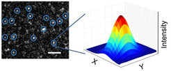
We use different forms of superresolution microscopy, such as Single Molecule Localization Microscopy (SMLM), STED microscopy or Airyscan, to study the nanoscale organization of macromolecular complexes in membranes. To improve data analysis, we developed a new software based on machine learning that allows the automatic detection, classification and quantitative description of supramolecular assemblies with distinct shapes (Danial & Garcia-Saez, Nat Methods, 2019). We aim to implement multi-color approaches as well as super-resolution live cell imaging to gain insight into the structural dynamics of death effectors.
With this approach, we were able to directly visualize the assembly of BAX oligomers into a distribution of lines, arcs and rings on apoptotic mitochondria (Salvador-Gallego et al., EMBO J, 2016).

2. Single Particle Stoichiometry Analysis

We use quantitative single particle imaging based on TIRF or confocal microscopy, in combination with fluorescence calibration standards, to determine the stochiometry of oligomeric complexes in membranes. Time-lapse analysis allows us to dissect their assembly dynamics.
This approach allowed us to find out that BAX exists in the membrane as a mixture of multiple oligomers based in dimer units (Subburaj et al., Nat Commun 2019).
3. Atomic Force Microscopy on Membranes

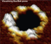
The use of AFM allowed us to visualize the membrane pores induced by BAX. In addition, force spectroscopy measurements provide information about the mechanical properties of the membranes and the alterations induced by cell death effectors.
4. Fluorescence Correlation Spectroscopy

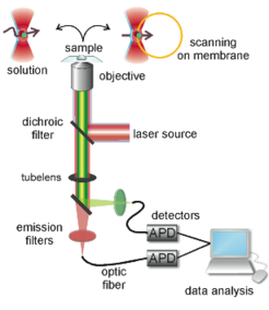
FCS is a technique with single molecule sensitivity that analyzes the fluctuations in fluorescence intensity within a tiny volume (in the order of fL). It can be used to measure diffusion coefficients, fluorophore concentrations, particle sizes, chemical reactions, conformational changes and binding/unbinding processes, among other. Two-color FCS or fluorescence cross-correlation spectroscopy (FCCS) is a variant of FCS extremely convenient for the study of molecular interactions, both in vitro and in vivo. It measures dynamic co-localization and therefore can be used for binding or dissociation studies. In the case of membrane applications of FCS, a very interesting approach is scanning FCS (SFCS).
Using this method, we quantified a minimal interactome composed of three representative BCL-2 proteins, BAX, cBID and BCL-xL, both in the membrane and in the cytosol, which allowed us to establish the hierarchy of interactions between the family members (Bleicken et al., Nat Commun, 2017).
5. Model Membranes
To investigate dynamic processes in biological membranes we use model systems of different complexity, ranging from pure lipid bilayers to cultured cells. A common feature of these membrane systems is that they can be visualized with optical microscopy and, as a consequence, they can be used for experiments of time-lapse microscopy, FRAP, FRET, FCS and other advanced microscopy techniques.

6. Optogenetic Control of Cell Death
We develop optogenetic systmes that allow the precise control of the activation of different cell death pathways at the single cell level with high temporal and spatial control. These new tools allow us to study the cross-talk between dying and bystander cells in vitro and in vivo, in order to understand how the different forms of cell death shapes tissue responses and the underlying mechanisms.


